
SPECIAL PROCEDURES
Extraction of all lipids with a mixture of hexane/isopropanol.
To reduce the danger of toxicity of chloroform, Hara et al. (Anal Biochem 1978, 90, 420) described an efficient extraction procedure particularly adapted to nervous tissues.
The tissue sample is homogenized with 18 volumes of a mixture of hexane/2-propanol (3/2) for 1 minute, the suspension is filtered and the filter rinsed with 3 x 2 vol of the same solvent. As the content of non-lipids is very low (proteins, pigments, small molecules), the whole liquid phase is evaporated and the dried extract dissolved (Eder K et al. Clin Chim Acta 1993, 219, 93).
An adaptation of the Hara’s method was demonstrated to be the most efficient procedure for the extraction of plant sphingolipids (Markham JE et al., J Biol Chem 2006, 281, 22684).
Briefly, to the frozen tissue, 5 ml of the lower phase of isopropanol/hexane/water, 55/20/25 (v/v) were added. The tissue was disrupted in a glass homogenizer and transferred to a glass tube which is capped and incubated at 60°C for 15 min with occasional shaking. After centrifugation, the pellet was extracted twice more, each time with 5 ml of solvent and the supernatants were combined. A 98% recovery was obtained with leaf tissue of Arabidopsis, tomato, and soybean.
An optimized procedure for extraction of total lipids from microalgae without disruption and using a chloroform/methanol mixture has been reported (Ryckebosch E et al., JAOCS 2012, 89, 189). The recovery of lipids from microalgae by alcohol processing was determined with two species, Nannochloropsis and Schizochytrium (Wang G et al., JAOCS 2012, 89, 335).
Alkanediol-based eutectic solvents have been proposed for extracting free fatty acids from Spirulina and Porphyridium Cakes (Wils L et al., Mar Drugs 2024, 22(6), 281).
Extraction of all lipids with methyl-tert-butyl ether (MTBE).
Accurate profiling of lipidomes were obtained by MTBE extraction which allows faster and cleaner lipid recovery (Matyash V et al., J Lipid Res 2010, 49, 1137). Because of MTBE’s low density, lipid-containing organic phase forms the upper layer during phase separation, which simplifies its collection and minimizes dripping losses. Nonextractable matrix forms a dense pellet at the bottom of the extraction tube and is easily removed by centrifugation. Rigorous testing demonstrated that the MTBE protocol delivers similar or better recoveries of species of most all major lipid classes compared with the Folch or Bligh and Dyer recipes.
Briefly, methanol (1.5 ml) was added to a 200 ml sample aliquot, and the tube was vortexed. Then, 5 ml of MTBE was added and the mixture was incubated for 1 h at room temperature. Phase separation was induced by adding 1.25 ml of water. Upon 10 min of incubation at room temperature, the sample was centrifuged at 1,000 g for 10 min. The upper organic phase was collected, and the lower phase was re-extracted with 2 ml of MTBE/methanol/water (10/3/2.5, v/v/v). Combined organic phases were dried in a vacuum centrifuge. Extracted lipids were dissolved in 200 ml of CHCl3/methanol/water (60/30/4.5, v/v/v) for storage.
A similar procedure has been described before analysis of blood lipid classes (Ichihara K et al., Lipids 2011, 46, 297). This extraction has been also proposed for the analysis of human brain lipids (Abbott SK et al., Lipids 2013, 48, 307). This study leads to the conclusion that this protocol, including a mechanical homogenization utilizing ceramic beads, is equivalent to the traditional Folch protocol for lipid extraction and quantification of glycerophospholipid, sphingolipid and sterol species in human brain tissue.
Extraction of all lipids with hexane
A very precise investigation of the effects of temperature and contact time on extraction efficiency of sunflower cake was reported using hexane as solvent (Baümler ER et al., JAOCS 2010, 87, 1489). Extraction at 60°C during 30 min leads to a very high yield (99%) for triacylglycerols and tocopherols and to a reduced phospholipid extraction (66%).
Extraction of lipids with 2-methyltetrahydrofurane (MeTHF), a bio-based solvent
Considering several theoretical and technical approaches, MeTHF (also named methyloxolane) can be considered as the best alternative to n-hexane among other tested solvents as it has good solubilization abilities regarding desirable compounds in oil (mainly glycerides). MeTHF is a highly flammable, mobile liquid but can be used as a replacement for tetrahydrofuran (THF) in several applications It is derived from sugars via furfural (it is a bio-based solvent)
The lipid yield and lipid profile of oils obtained is comparable to the one extracted with hexane extraction. No significant selectivity between n-hexane and MeTHF was noted as the composition in fatty acids remains the same. Therefore, the bio-based solvent MeTHF (or 2-methyloxolane) can be considered as an excellent alternative to the petroleum-based solvent n-hexane for edible/cosmetic oil extraction (Gharby S et al., OCL 2020, 27, 27). Its utilization for oil extraction can drastically reduce the health and environmental impacts associated with n-hexane. Authors have used a soxhlet system to extract the lipids from vegetal sources.
Economic and energetic evaluations of the process were conducted to estimate that bio-based MeTHF is a valuable alternative solvent compared to hexane as petroleum solvent for extraction of fats and oils (Sicaire AG et al., Int J Mol Sci 2015, 16, 8430). Further comparative results concerning aroma, carotenoids, fatty acids, glycerides, sterols and phospholipids have been reported (Rapinel V et al., Molecules. 2020, 25, 3417).
A study has compared the effects 2-methyltetrahydrofuran on oil yield, stability and bioactive compounds. MeTHF increased oil yield from 12 to 28 %) (Erdoğan U et al., Food Chem 2025, 478, 143659). Solvent type did not change fatty acid and tocopherol composition. MeTHF increased number of phenolic compounds from 9 to 16 and amount of total tocopherol, phenolics, chlorophylls and carotenoids in oils. MeTHF extracted oils had significantly higher antioxidant capacity.
The concept of green solvent and the properties and characteristics to be considered green have been reviewed (Calvo-Flores FG et al., Top Curr Chem 2018, 376:18).
Extraction of dry tissue lipids
Based on precise studies, it has been shown that freeze-dried tissues (krill) extracted with a one-step procedure (acetone:ethanol, 1:1, v:v) using 1:12 krill:solvent ratio (w:v) resulted in the highest oil extraction efficiency (Gigliotti JC et al., Food Chem 2011, 125, 1028). That oil contained about 30% of phospholipids, 3% of triacylglycerols and 67% of polar non-phospholipid classes (cholesterol, mono- and diacylglycerols, astaxanthin).
Dry-column method
An alternative to the traditional Folch method was described using solvent elution of a dry column composed of a tissue sample, anhydrous sodium sulfate, and Celite diatomaceous earth ground together (Marmer WN et al. Lipids 1981, 16, 365). Alternatively, lipids may be isolated and simultaneously separated into neutral and polar fractions by a sequential elution procedure. Analyses of muscle and adipose tissues demonstrated that results were similar to those obtained with chloroform/methanol methods.
A less time-consuming dry-column method was scaled down and adapted to 1g samples (liver and muscle tissues) (Elmer-Frohlich K et al., JAOCS 1992, 69, 243).
Procedure: The sample (about 1 g) is ground for 30 sec. in an ice-chilled mortar with 4 g anhydrous Na2SO4 and 0.1 ml BHT (20 mg/l dichloromethane). Celite 545 (3 g) (Fisher Scientific) is added and the mixture is ground for an additional 30 sec. to obtain a fine homogenized powder.
The powder is poured into a 16 mm X 30 cm glass column packed with glass wool and 2g of CaHPO4/Celite 545 (1/9 w/w) at its tip. A slight compression was accomplished at the top with a glass rod. Mortar, pestle, and glass rod are rinsed with 15 ml of dichloromethane/methanol (9/1, v/v) which are transferred into the column. In addition to these 15 ml, 50 ml of solvent mixture are added to elute lipids which are isolated, weighed and analyzed after evaporation of the solvent under nitrogen flushing.
Multiple columns may be run simultaneously.
A patented process, first reported in 1989 (Barker SA et al., J Chromatogr 1989, 475, 353), known as “matrix solid-phase dispersion” was described to conduct simultaneously disruption and extraction of solid and semi-solid samples. Thus, a highly viscous, semi-solid or solid sample can be placed in a mortar containing a bonded-phase solid support material (C18 bonded silica) and mechanically blended to perform a complete disruption and dispersal of the sample. This blend is sufficiently dry to transfer and pack into a column for more classical application of solid-phase extraction to the isolation of sample components. This technique has been most frequently applied to the isolation of drugs, herbicides, pesticides and other pollutants from animal tissues, fruits and vegetables (review in : Barker SA, J Chromatogr A 2000, 885, 115).
Bligh and Dyer Method
Bligh and Dyer (Can J Biochem Physiol 1959, 37, 911) introduced a method where extraction and partitioning are simultaneous, the precipitated proteins are isolated between the two liquid phases. This method is particularly suitable for lipid extraction of incubation medium, tissue homogenates or cell suspensions. The extraction can be carried out in a single tube where previous studies took place.
It must be pointed out that this extraction method was shown to give significantly lower estimates of lipid content in samples containing more than 2% lipid (mainly triacylglycerols) and this underestimation increased with increasing lipid content of the sample (Iverson SJ et al. Lipids 2001, 36, 1283). Thus, the total lipid content of fatty samples are accurately determined using the Folch extraction method.
Procedure: To a sample containing 1 ml water (1 ml cell suspension, homogenized tissue, plasma…), add 3.75 ml of a mixture chloroform/methanol (1/2) and vortex during 10-15 min, then add 1.25 ml chloroform with mixing 1 min and 1.25 ml water with mixing another minute before centrifugation. Discard the upper phase and collect the lower phase through the protein disk with a Pasteur pipette. For large volumes of liquid, it is advisable to filter the mixture to remove the insoluble parts of the sample and to centrifuge the liquid phase to allow the formation of the two liquid phases. After evaporation, the lipid extract (lower phase) will be redissolved in a small volume of chloroform/methanol (2/1).
The basic procedure was improved to increase the yield of lipids. One of the most common modifications is to replace water by 1M NaCl. This addition blocked the binding of some acidic lipids to denatured lipids. If necessary, the addition of 0.2 M phosphoric acid to the salt solution is possible (Hajra, lipids, 1974, 9, 502) to improve their recovery. In this case, plasmalogens are converted to lyso lipids.
If an exhaustive extraction is necessary, an extraction with two steps can be used.
Similarly, it was described that the addition of acetic acid (0.5% v/v) in the water phase significantly increased the recovery of acidic phospholipids (Weerheim AM et al., Anal Biochem 2002, 302, 191). Another modification has been proposed, in comparison with the traditional extraction method (Jensen SK, Lipid Technol 2008, 20, 280). Acidification with HCl improved the extraction of lipids in a shorter time.
Two-step Bligh and Dyer method
To a sample containing 1 ml water (1.25 g tissue, 1 ml cell suspension, homogenized tissue, plasma…), add 3.75 ml of a mixture chloroform/methanol (1/2) and vortex during 10-15 min, then add 1.25 ml chloroform with mixing 1 min and 1.25 ml water with mixing another minute before centrifugation. Collect the lower phase in another glass tube.
Add 1.88 ml of chloroform to the non-lipid residue, vortex, centrifuge. Mix the lower phase to the first chloroform phase in the glass tube. After evaporation, the lipid extract will be dissolved in a small volume of chloroform/methanol (2/1).
Extraction of bacteria
An efficient modification of the Bligh and Dyer method, given below, was proposed for the extraction of lipids from unicellular organisms. Several parameters were optimized to improve the fatty acid recovery (Lewis T et al., J Microbiol Meth 2000, 43, 107). Thus, it was shown that the total amount of recovered fatty acids increased by about 30% by adding solvents to the biomass in order of increasing, as opposed to decreasing, polarity.
Cells were harvested by centrifugation at high speed for 15 min. The supernatant was discarded, the cell pellet re-suspended in 100 ml 1.0% NaCl (w/v), and re-centrifuged. The second supernatant was discarded and the cell pellet frozen overnight at -30°C. Frozen biomass was freeze dried for 15 h and subsequently stored in a sealed glass container at -30°C.
To freeze-dried cells (about 100 mg) to which a total of 114 ml solvents were added in the sequence: chloroform, methanol, water to achieve a final chloroform/methanol/water ratio of 1/2/0.8 (v/v/v). Samples were shaken for 15 s immediately following the addition of each solvent, and allowed to stand for about 18 h, with occasional shaking by hand.
Phase separation of the biomass-solvent mixtures in the separatory funnels was achieved by adding chloroform and water to obtain a final chloroform/methanol/water ratio of 1/1/0.9 (v/v/v). A known portion of each total lipid extract recovered from the lower chloroform phase was used for further analysis.
Comparing various extraction procedures, it has been shown that a modified (miniaturized) Bligh and Dyer extraction technique was the most efficient with an oleaginous bacteria Thraustochytrium sp (Burja AM et al., J Agric Food Chem 2007, 55, 4795). If only fatty acid determination is required, a direct saponification using KOH in ethanol was almost as efficient as the previous one.
An hexane-isopropanol solvent system was found to be effective for the bacterium Pseudomonas atlantica (Guckert J B et al., J Microbiol Methods 1988, 8, 131), this system gaving a lower recovery of lipids in the green alga Chlorella sp. (Guckert J B et al., J Microbiol Methods 1988, 8, 139).
At work using a yeast, Yarrowia lipolytica, has reported that bio-based solvents could be an alternative to petrochemical solvents, such as hexane. Some differences were noted between experimental and theoretical studies (Breil C et al., Molecules 2016, 21, 196). Ethyl acetate and methyltetrahydrofuran are the best candidate solvents to extract all of the lipids of Yarrowia lipolytica (triglycerides, diglycerides, free fatty acids and
phospholipids) and are derived from renewable resources.
Extraction of highly polar lipids
When tissues are rich in highly polar lipids such as gangliosides, a reliable extraction method is needed to prevent their loss while extracting all other lipid classes. All the previously described techniques brings gangliosides and likely a part of other very polar lipids into the water-rich layer. A method, recently described for nervous tissues (Dreyfus et al., Anal Biochem 1977, 249, 67-78), prevents these drawbacks.
Small tissue samples corresponding up to 10 mg protein suspended in 0.5 ml water are mixed with 5 ml chloroform/methanol mixture (1/1) for 30 min. The pellet obtained by centrifugation is extracted again with successively 3 ml of the same solvent, 3 ml of a mixture chloroform/methanol (1/2) and 3 ml of a mixture chloroform/methanol/water (60/30/4.5). The four lipid extracts are combined and evaporated and the dry residue dissolved for further purification.
A liquid/liquid extraction has been used with success for mass spectroscopy estimation of brain gangliosides (Garcia AD et al., J Chromatogr B 2014, 947-8, 1). The lipid extract was suspended in a mixture of chloroform/methanol/water (30/60/8, v/v/v), vortexed and sonicated. The solution was then centrifuged and the aqueous phase containing the enriched gangliosides was collected and set aside while the organic phase was then subjected to the same extraction procedure. The supernatants were combined and dried under vacuum. Once dry, the lipid extracts were resuspended in 10 mL of the liquid chromatography starting buffer. The gangliosides were purified on C8 SPE cartridges before LC-MS analysis.
A very efficient method has been developed to isolate and purify GM1 from pig brain (Bian L et al., Biomed Chromatogr 2015, 29, 1604). The method consisted of a precipitation by acetone followed by an extraction by chloroform–methanol–water. The purification was done using a two-step chromatographic separation by DEAE–Sepharose Fast Flow anion-exchange medium and Sephacryl S-100 HR size-exclusion medium. The final yield of GM1 was about 0.022% ( g/g) with the purity of about 98%. This method will probably provide a reference alternative for isolation and purification of other amphipathic substances in biological tissues.
Lyso derivatives (N-deacylated) of glycosphingolipids are not efficiently recovered from cell extracts because of their high polarity. The glycosylsphingosine are found in tissues from patients and in animal models. In normal conditions, their concentration are very low and needs very efficient techniques to be evaluated. It was shown that a second extraction with water-saturated butanol of the upper aqueous phase obtained after a Folch extraction was necessary to recover up to 98% of the tissue lysoglycosphingolipids (Bodennec J et al., J Lipid Res 2003, 44, 218).
Extraction of plasma total lipids
For the extraction of plasma lipids we used a very rapid and efficient method.
To 0.2 ml plasma add 0.3 ml 0.5 M KH2PO4 , 1.5 ml chloroform and 0.5 ml methanol. After vortexing 2 minutes and centrifugation, the lower phase is collected with a Pasteur pipette through the protein disk and evaporated.
A procedure using the detergent Triton X-114 was shown to be very efficient for the extraction of plasma lipids, while sparing the protein fraction for further use (Ferraz TPL et al., J Biochem Biophys Meth 2004, 58, 187).
A method based on the modification of an extraction method originally developed for pesticide residue analysis in food has been described for the purpose of isolating lipids from biological fluids (plasma, urine) (Bang DY et al., J Chromatogr A 2014, 1331, 19). The procedure adapted for the preparation of samples for mass speectrometry includes a extraction/partitioning step with a mixture of CHCl3/CH3OH in the presence of MgSO4 and CH3COONa and an adsorption/desorption step with C18 particles. That method was applied to lipid extracts from both human urine and plasma samples, demonstrating that it can be powerfullyutilized for high-speed (<15 min) preparation of lipids compared to the Folch method, with equivalent or slightly improved results in lipid identification using liquid chromatography and mass spectrometry.
A fast, simple extraction method with high recovery (>90%) and reproducibility (%CV < 20%) has been described (Alshehry ZH et al., Metabolites 2015, 5(2), 389). It showed a strong correlation of all lipid measures with an established chloroform:methanol extraction method. This method uses non-halogenated solvents, requires no drying or reconstitution steps and is suitable for large-scale LC ESI-MS/MS-based lipidomic analyses in research and clinical laboratories. Plasma (10 μL) was mixed with 100 μL 1-butanol:methanol (1:1 v/v) containing internal standards resulting in efficient extraction of all major lipid classes (including sterols, glycerolipids, glycerophospholipids and sphingolipids).
Delipidation of plasma, serum or plant seeds
When plasma proteins, including the apolipoproteins, must be preserved from denaturation during the extraction of lipids a specific solvent system must be used (Cham BE et al., J Lipid Res 1976, 17, 176).
The most common procedure used for delipidation of plasma, protein solutions or cell culture medium involves the extraction of all kinds of lipids with a mixture of butanol and di-isopropyl ether. The proteins remain in solution in the aqueous phase, while the organic phase contains the dissolved lipids.
Procedure : One volume of serum or plasma containing 0.1 mg/ml of ethylenediamine tetraacetate (EDTA) is added to 2 volumes of a mixture of butanol/di-isopropyl ether (40/60, v/v). The vials are tightly closed and fastened on a mechanical rotator providing end-over-end rotation at about 30 rpm for 0.5 h.
After extraction, the mixture is centrifuged at low speed (2000 rpm) for 2 min to separate the aqueous and organic phase. The aqueous phase containing the delipidated proteins is removed by careful suction with needle and pump or syringe.
Traces of butanol remaining in the aqueous solution may be removed if necessary by washing that phase with 2 volumes of di-isopropyl ether. Residual solvent may be removed by an extraction with a water pump aspirator at 37°C for some minutes.
When proteins from plant oilseeds are studied by electrophoresis, lipids must be removed to prevent important interferences and thus to obtain good resolution. It was demonstrated that, in the presence of chloroform methanol, lipid contaminants can be thoroughly removed by the combination of two precipitation steps (10% TCA/acetone and acetone) and aqueous TCA wash steps (Wang W et al., Anal Biochem 2004, 329, 139).
Extraction of plasma fatty acids and acylglycerols
A reliable extraction procedure was described by Dole (J Clin Invest 1956, 37, 350) for the extraction of free fatty acids and non polar acylglycerols.
To 1 ml plasma (or aqueous solution) add 5 ml of Dole reagent (40 ml isopropanol, 10 ml heptane and 1 ml 1M H2SO4), 3 ml heptane and 2 ml water.
After vortexing and centrifugation, the upper phase is collected. Residual phospholipids may be removed by the addition of 200 mg silica gel (vortex and centrifuge) before evaporation under nitrogen.
Phosphoinositides extraction
Several procedures were described to extract the polyphosphoinositides since they are known to bind strongly to proteins during their denaturation. To improve the recovery, the use of an acidic solution is necessary. We used the procedure of Honeyman (Biochem J 1983, 212, 489) which is a modification of the Lloyd’s method (Br J Haematol 1972, 23, 571-585).
This is the one-step method of Bligh and Dyer modified by the inclusion of HCl to improve recovery of acidic phospholipids :
1 ml of cell suspension is mixed with 3.75 ml of chloroform/methanol/12N HCl (2/4/0.1, v/v). After thorough mixing, 1.25 ml of chloroform is added with vortexing 30 sec followed by 1.25 ml of water with similar mixing. After centrifugation 10 min at low speed, the lower chloroform layer is removed and transferred to a glass tube for evaporation.
Method used to extract platelet phosphoinositides
Stop incubation of the cell suspension by adding 1 ml of chloroform/methanol (1/1) to 1.5 ml of aqueous medium and vortex some seconds. Transfer the mixture with a Pasteur pipette into a 15 ml Falcon polypropylene tube.
Add to the mixture 4 ml of chloroform/methanol (1/1), then 0.4 ml 10N HCl and then 0.5 ml water. Vortex during 5 min to extract lipids and centrifuge the plastic tube at low speed 10 min at 4°C.
Transfer the lower phase into a second Falcon tube by sampling through the proteinaceous disk with a Pasteur pipette. Add to this solution 2.5 ml methanol, 2.1 ml water and 0.4 ml 10N HCl. Vortex some seconds and centrifuge at low speed in the cold.
Transfer the lower phase as previously in a glass tube, evaporate with the help of nitrogen and dissolve the residue with a small volume of chloroform/methanol (1/1).
Extraction of plant material
Plant tissues are difficult to extract because of active lipases which hydrolyze rapidly phospholipids glycolipids and increase the amount of free fatty acids in the extract. Thus, a solvent frequently used to inhibit these enzymes is isopropanol.
Nichols’ method: Plant tissues are minced and macerated with 100 parts (w/w) of isopropanol. The mixture is filtered, the solid is extracted again with 200 parts of chloroform/isopropanol mixture (1/1, v/v). The combined filtrates are evaporated, dissolved in a small volume of chloroform/methanol (2/1, v/v) and, if necessary, washed according the Folch’s procedure. Optimal conditions of extraction of microalgae with aqueous isopropanol combined or not with cell rupture has been reported (Yao L et al., JAOCS 2013, 90, 571). The oil yield was largely increased after ultrasonic cell rupture after extraction with 88 or 95% isopropanol.
The extraction of algae is made with hot isopropanol (60°C) added to the cell suspension. It appears that unicellular algae (plankton) must be extracted rapidly with a minimum of preparative mechanical treatments (centrifugation, filtration). A preliminary small scale extraction is recommended to choose the procedure to be adopted.
A comparative evaluation of several extraction methods was done in three types of macroalgae (Kumari P et al., Anal Biochem 2011, 415, 134). Care should be taken while selecting the method for macroalgae, according to the group to which they belong, otherwise there would be a risk of obtaining erratic and inaccurate results.
The extraction of neutral lipids from microalgae has been efficiently done on lyophilized material (Fajardo AR et al., Eur J Lipid Sci Technol 2007, 109, 120). Briefly, first, 96% ethanol was used to extract the lipids from the dry biomass. Second, a biphasic system was formed by adding water and hexane to the extracted crude oil. Thus, the purified lipids were transferred to the hexane phase while most impurities remained in the aqueous phase.
An overview of advances made in technologies for extracting microalgae oil may be consulted before doing experiments (Mercer P et al., Eur J Lipid Sci Technol 2011, 113, 539). Solvent extraction technologies with extraction alternatives such as mechanical milling and pressing, enzymatic and supercritical fluid extraction are compared.
A comparative study of various extraction treatments have been reported using the microalgae Spirulina (Zheng G et al., JAOCS 2012, 89, 561). A purification was used to remove the pigments from the extracted lipids.
Efficient extraction of carotenoids from dry plant material (maize endosperm) has been described using mixtures of methanol/ethyl acetate (6/4, v/v) and methanol/tetrahydrofuran (1/1, v/v) (Rivera S et al., Molecules 2012, 17, 11255).
Extraction of plant sphingolipids
Because of their large hydrophilic polar head, solubilisation of glycosylated sphingolipids in usual organic solvents is inaccurate. When doing a phase partition in chloroform/methanol/water mixtures, these lipids remain insoluble for the most part or are recovered in the aqueous phase and interphase. A new extraction method to purify total plant sphingolipids has been developed (Buré C et al., Rapid Commun. Mass Spectrom 2013, 25, 3131). Briefly, plants cells were blended with cold 0.1 N aqueous acetic acid in a chilled Waring Blendor. The slurry was filtered under vacuum and the aqueous acetic acid filtrate was discarded. The residue was then re-extracted with hot 70% ethanol containing 0.1 N HCl. The filtrate was chilled and left at room temperature overnight. The precipitate was pelleted by centrifugation. The sphingolipid-enriched pellet was washed twice with cold acetone, and twice with cold diethyl ether to yield a whitish precipitate. Glycosylated sphingolipid contained in the precipitate were then dissolved in tetrahydrofuran (THF)/methanol/water (4/4/1, v/v/v) containing 0.1% formic acid by heating at 60°C, followed by gentle sonication. This solution was further used for mass spectrometry analyses (Buré C et al., Rapid Commun Mass Spectrom 2011, 25, 3131; Cacas JL et al., Phytochemistry 2013, 96, 191).
Enzyme-assisted aqueous extraction (EAEP)
EAEP has been employed to extract different compounds from plants, and has been proved to be effective in improving the yield of various components. Improved lipid extraction was observed in many different oil-bearing plant materials including soybean (Freitas SP et al., Fett-Lipid 1997, 99, 333), sunflower seeds (Sineiro J et al., Food Chem 1998, 61, 467) and sesame (Latif S et al., Food Chem. 2011, 125, 679).14 In addition, EAEP will make it possible to extract and separate oil directly from algae in the natural aqueous environment of algae cultures, which avoids the collection and drying process of algae biomass.
As insoluble nonhydrolyzable biopolymers (algaenans) are present in cell wall of algae, the EAEP methods established for common terrestrial plants cannot be applied directly to the lipid extraction from microalgae. Improvement in lipid extraction of these vegetals has been done in using sonication combined with enzyme treatment (Liang K et al., J Agric Food Chem 2012, 60, 11771).
Oilseeds extraction
They may be analyzed for oil content by an exhaustive extraction with petroleum ether. A comparison of five methods to measure the oil contents in oilseeds may be studied before choosing a specific procedure (Barthet VJ et al., J Oleo Sci 2002, 51, 589).
A one-tep extraction has been described to study the composition of triglycerides in small piece of seed and is suitable for a large number of tissue samples should be examined as in selecting new plant varieties. This easy and reliable method is based on an incubation of samples (20-50 mg) without shaking in a mixture containing heptane / 0.17 M NaCl in methanol (66.6/33.3, v/v), for 2 h at 80°C. After cooling, the upper phase containing mainly triglycerides was transferred to a new test tube for further analysis. Even under incomplete triglyceride extraction (80% maximum) the triglyceride ecomposition is representative of the total triglycerides found in the tissue (Ruiz-Lopez N et al., Anal Biochem 2003, 317, 247).
2-Methyltetrahydrofuran was successfully proposed instead of petroleum solvents for the extraction of fat and oils for food (edible oil) and non-food (bio fuel) applications (Sicaire AG et al., Int J Mol Sci 2015, 16, 8430). Experiments were performed on rapeseed oil extraction at laboratory and pilot plant scale for the determination of lipid yields (glycerides, fatty acids, phospholipids, tocopherol, sterols).
An efficient surfactant-based extraction of corn oil from corn germ has been proposed (Kadioglu SI et al., JAOCS 2011, 88, 863). Hexane and/or other organic solvents were avoided in the process. Greater than 80% of corn oil can be extracted with low surfactant and salt concentrations. It was concluded that aqueous-based surfactant microemulsion oilseed extraction is a promising alternative approach for oil extraction.
The extraction of xanthophyll was shown to be improved using cellulolytic enzymes and highly competitive when compared to the traditional process of pigment extraction (Navarrete-Bolanos JL et al., J Agric Food Chem 2004, 52, 3394). These data may foster the development of new extraction procedures in plants based on previous enzymatic hydrolysis of cell membranes.
The extraction of flour can be made with hexane at room temperature but the efficiency of phospholipid recovery is dependent upon the temperature and the moisture content (Snyder HE, Inform 2004, 15, 575).
The reading of the review of various techniques of preparing plant material by Romanik G et al. is suggested before any plant extraction (Romanik G et al., J Biochem Biophys Methods 2007, 70, 253).
Extraction of essential oils
Hydrodistillation with electric heating is commonly used as part of the extraction. The Clevenger apparatus is used to collect evaporating essential oils. This method is environmentally friendly and solvent-free, ensuring the purity of the extract. Microwave was used to increase the efficiency of the distillation.
The sample is boiled in the boiling flask, steam rises in the assembly up to a condenser, and the condensate falls into the small extractor burette (water is returned to the tank through a channel whose position differs according to the densitiy of the oil in relation to water). After some time, the oil volume collected in the burette can be directly measured.
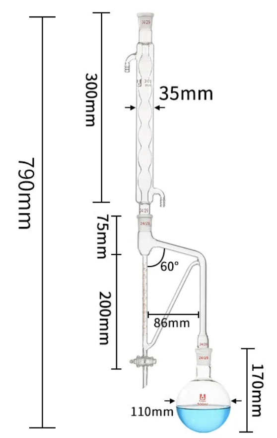 Clevenger hydrodistillation device
Small scale oil production
We describe here the extraction on a laboratory scale of lipids from various biological materials to run analytical procedures. Different processes may lead also to the extraction of oil from many seeds, nuts, and kernels for use as nutritional supplement, as well as raw material for industrial applications.
The type of instrument that is appropriate depends on the size of the operation. Oil processing operations range from “cottage industries” processing some kg per day, to factories processing several thousand tons of seed per day. In all cases, the sequence of operations includes cleaning, dehulling, grinding, and pressing. Furthermore, oils generally need refining to remove cloudiness, excess color and unpleasant flavors. As many crude oils contain variable amounts of mucilaginous compounds (gums) combined with phospholipids, a degumming process has to be run (Dijkstra AJ, OCL 1998, 5, 367). In several instances, and especially when seeds have low oil content (like soybeans) more complex methods including solvent extraction are processed. If hexane is the most commonly used solvent, isohexane appears to be the most likely candidate to replace n-hexane as the preferred oilseed extraction solvent, mainly in USA to avoid federal environmental regulations applying to the use of n-hexane. Several studies have shown that the extraction performances of the two isomers are quite similar (Inform 2002, 13, 282).
Those who are interested in oilseed processing will find a comprehensive description of basic processes with sources for additional information and a list of suitable raw material in the ATTRA (National Sustainable Agriculture Information Service) publication :
Small-scale oilseed processing. Value-added and processing guide
Aqueous enzymatic oil extraction is an emerging technology since it offers many advantages such as elimination of solvent consumption, degumming operations and removal of some toxins. A review of aqueous and enzyme-based processes may be consulted (Rosenthal A et al., Enzyme Microb Technol 1996, 19, 402). Applications of that methodology to the oil extraction of Irvangiaseed kernels (Women HM et al., Eur J Lipid Sci technol 2008, 110, 232) and canola seeds (Latif S et al., Eur J Lipid Sci Technol 2008, 110, 887) have been reported.
Comparative experiments on tobacco seeds have demonstrated that the best oil recovery was obtained by cold pressing (Stanisavljevic IT et al., Eur J Lipid Sci Technol 2009, 111, 513). Shortly, seeds were pressed by a hydraulic press (Komet, Germany) through three nozzles (diameter of 15, 10 and 6 mm). The press does not involve mixing and tearing of the seeds. The seed cake was ground by an electrical mill and the residual oil was extracted by using n-hexane at a seeds-to-solvent ratio of 1 : 3 g/mL and 25°C for 60 min. This recovery technique is said to be more acceptable than the other methods, not only for economic but also for environmental, health and safety reasons.
As hexane presents numerous drawbacks like high flammability, dangerousness for health and environment, it has ben variously replaced by agro-solvents. An overview of these techniques are presented in the work of Fine F et al. (Fine F et al., OCL 2013, 20, A502).
Very small samples
When tissue samples are very tiny (1 mm or less), the analysis of their lipid content (neutral lipids or phospholipids) may be effected directly on the TLC plate without previous extraction (Lecomte M et al., Prostagl Leukot Essent Fatty Acids 1998, 59, 363).
Procedure:
The tiny piece of tissue is directly applied in a small hole or groove made in the concentration zone (kieselgur preadsorbent) of the TLC plate (cf Whatman plates). This plate is then rapidly applied onto a steel plate pre-cooled in liquid nitrogen. After one or two minute freezing, the plate is lyophilized one or two hours under vacuum. The plate is then rapidly submitted to a solvent elution adapted to the desired analysis.
Mineralized samples
As some acidic phospholipids (mainly phosphatidylserine) are known to complex with calcium, particularly in the presence of Pi, a complete extraction of membrane lipids in mineralized tissues must be done after chemical demineralization (Wu L et al., J Biol Chem 2002, 277, 5126). This approach was first discovered by Shapiro IM in 1971 (Arch oral Biol 1971, 16, 411; Calcif Tissue Res 1970, 5, 21). A complete extraction and analysis of mineralized bone tissue and bone marrow lipids have been described in detail (During A, J Chromatogr A 2017, 1515, 232).
Procedure:
Small tissue samples are powdered in the cold (Freezer mill) and extracted with chloroform/methanol (2/1) mixture. After a short sonication (1-2 min), the suspension is centrifuged (3000 rpm, 12 min) and the extract collected. The pellet is demineralized with 0.5M Na salt EDTA for 20-30 min and sedimented after a short centrifugation. After removal of the supernatant, the decalcified residue (pellet) is reextracted using a chloroform/methanol/conc. HCl (200/100/1) mixture. The two lipid extracts are mixed, dried and washed with saline to remove non-lipid contaminants.
Skin surface
Several lipids are present at the surface of mammalian skin. Their analysis is of great interest in relation with medical treatment in diseases such as acne, atopic dermatitis, seborrhea or psoriasis. Applications may be extended to cosmetic and alimentary fields. The extraction of surface lipids is efficiently processed with a very simple design (Michael-Jubeli R et al., J Lipid Res 2011, 52, 143).
Briefly, two lipid-free absorbent papers are maintained on the defined area for 30 minutes using a medical tape, and then removed with tweezers and introduced into a closed vial. This step was repeated four times. The collected lipids are extracted from papers twice with 40 ml of diethyl ether. The solution is concentrated and transferred into a vial.
A novel method for the extraction of insect cuticular hydrocarbons has been described (Choe DH et al., J Chem Ecol 2012, 38, 176). The cuticular hydrocarbons are first adsorbed to solica gel and then are eluted using organic solvents.
DISPERSIVE LIQUID-LIQUID MICROEXTRACTION
Lire la suiteDevenez membre et participez au développement de la Lipidomique au XXIème siècle.
S'inscrire