
VITAMIN K
The vitamin K family is comprised of multiple similarly structured fat-soluble molecules containing a 2-methyl-1,4-naphthoquinone ring structure called menadione. Menadione (K3) is of synthetic origin, though due to reported adverse effects of hemolysis and liver toxicity will not encompass the scope of this page.
Naturally, vitamin K occurs as two vitamers: vitamin K1 (also known as phylloquinone) and vitamin K2 (designated also as menaquinones (MKs).
Differences between isoforms vitamin K1 and K2 by means of source, function, and extrahepatic activity have been reported (Halder M. et al., Int J Mol Sci 2019, 20, E896).
The existence of the vitamin K group was discovered by H. Dam (Biochim. Zeitschr. 1929, 215, 475) in studying cholesterol metabolism in chicks. He noted a new deficiency syndrome in the young birds fed a fat deficient diet. The characteristic features were a lengthened blood clotting time, anemia and hemorrhage. Ten years later, E.A. Doisy‘s group (Binkley et al., J. Biol. Chem. 1939, 130, 219) succeeded in isolating the vitamin from hexane extracts. Although at the time the structure had not been established, chemical and physical properties were correctly attributed to a substituted 1,4-naphthoquinone. Subsequent works by H. Dam, rewarded with the Nobel prize for medicine in 1943 “for his discovery of vitamin K”, with E.A. Doisy “for his discovery of the chemical nature of vitamin K”, led to the characterization of the molecule he termed vitamin K (Koagulationsvitamin).
Vitamin K1 was named phylloquinone since it is an indirect product of photosynthesis in plant leaves where it occurs in chloroplasts and participates in the overall photosynthetic process. The methyl naphthoquinone ring (chromene) has a phytyl side chain (partially saturated poly-isoprenoid alcohol).
Vitamin K (its reduced form) is necessary for post-translational modification of coagulation factors II (prothrombin), VII, IX and X in the liver. In the presence of the reduced form of vitamin K (dihydro vit K or vitamin K hydroquinone) formed by the action of a reductase enzyme on phylloquinone, certain glutamic acid residues of the nascent polypeptides are converted to γ-carboxyglutamic acid by a carboxylase enzyme. This confers the ability to bind calcium, a property essential for the physiological function of these proteins. Vitamin K has been investigated for its role in altering bone metabolism. Its impact on bone health involves its absorption, metabolism and excretion, while the potential mechanisms associated with its effect on bone metabolism include its action on vitamin K-dependent proteins to increase deposition and bone formation through various pathways, as recently reviewed (Wang H et al., Nutrients 2023, 15, 4935).
Vitamin K is also involved in the activation of a protein, the matrix Gla protein (MGP), an inhibitory factor which is highly expressed by vascular smooth muscle cells and regulates vascular calcification (Proufoot D et al., Nephrology 2006, 11, 455). The role of vitamin K and the potential antagonism by anticoagulants have been reviewed on the basis of bone health and osteroporosis (Pearson DA, Nutr Clin Pract 2007, 22, 517). Evidence is mounting that vitamin K1 may play an important role in the visual system apart from the hepatic carboxylation of hemostatic-related proteins. A review is covering that topic (Mong MA, Nutrients 2023, 15, 1948).
Vitamin K1 has nothing to do in plants with blood clotting, or even amino acid modification. It has been demonstrated that it acts to move electrons in the photosynthetic machinery (Hauska G, Trends Biochem Sci 1988, 13, 415). At some point in evolution, animals have reused the molecule into a completely different metabolism.
During the vitamin K action, a molecule of vitamin K epoxide (vit K 2,3-oxide) is formed which is further reduced, mainly by cellular thiol reagents, back to vitamin K1 (phylloquinone).
Vitamin K1 (MW: 450.7) is a yellow viscous oil with a maximum absorbance at 248 nm (molar abs coeff.:19900, the same for menaquinones), soluble in ethanol, hexane, chloroform and vegetable oils. Vit K1 is sensitive to sunlight (destroyed after one hour), unaffected by diluted acids but destroyed by basic solution and transformed by reducing agents. Vitamin K1 oxide (MW: 466.7) is much more stable to light and soluble in the same solvents as Vitamin K1. It has a maximum absorbance at 259 nm (molar abs. coeff.: 6170).
The K vitamins are subject to side-chain structural isomerization, and naturally occurring K1 is found exclusively as the biologically active 2′-trans-isomer. However, biological samples may contain some quantities of the inactive cis-isomer. Thus, its amount must be estimated in establishing reliable food databases.
| Food | Concentration (μg/100 g) |
| kale | 800 |
| spinach | 380 |
| brocoli | 240 |
| Brussel sprout | 180 |
| cabbage | 145 |
| asparagus | 100 |
| peas | 40 |
| avocado | 40 |
| lettuce | 35 |
| kiwi | 25 |
| soybean oil | 193 |
| rapeseed oil | 127 |
| cottonseed oil | 60 |
| olive oil | 55 |
Phylloquinone is poorly represented in fruits (0.1 to 3 μg per 100 g) except avocado (40 μg per 100 g) and kiwi (30-50 μg per 100 g), blueberries (15–27 μg per 100 g), blackberries (15–25 ug per 100 g), grapes red and green (14–18 μg per 100 g), dried figs (11–20 μg per 100 g) and dried prunes (51–68 μg per 100 g). K1 is present in several nuts : pine nuts (33–74 μg per 100 g), cashews (19–64 μg per 100 g), and pistachios (10–15 μg per 100 g)
Grain products have also very low levels of vitamin K1 (0.1 to 7 μg per 100 g) . Animal products including eggs do not appear to contain appreciable amounts of vitamin K1 (less than 50 ng/g) and less than 1 μg per 100 g are found in fish and shellfish. High amounts are found in butter (up to 100 μg per 100 g) but lower amounts in cheese (2-10 μg per 100 g). In contrast, a high diversity of menaquinones are present in dairy products.
Kiwi fruit (33.9–50.3 μg per 100 g), avocado (15.7–27.0 μg per 100 g), blueberries (14.7–27.2 μg per 100 g), blackberries (14.7–25.1 ug per 100 g), grapes red and green (13.8–18.1 μg per 100 g), dried figs (11.4–20.0 μg per 100 g) and dried prunes (51.1–68.1 μg per 100 g). K1 was present in several nuts during this study; pine nuts (33.4–73.7 μg per 100 g), cashews (19.4–64.3 μg per 100 g), and pistachios (10.1–15.1 μg per 100 g)
In animal tissues, phylloquinone is distributed in liver but also in heart, but is present at low concentrations in brain (Thijssen H et al., J Nutr 1996, 126, 537).
An adequate intake for a 25-year old male for Vitamin K is about 120 micrograms/day.
![]()
A second vitamin K (vit K2) was isolated very early from putrefied fish meal as a product of microbial synthesis (McRee RW et al., J. Am. Chem. Soc. 1939, 61, 1295). It has the same absorption peaks in UV region as vitamin K1 but with a slightly lower intensity (molar abs. coeff. at 248 nm: 11800). Vitamin K2 has a poly-isoprenoid unsaturated side-chain of various length, with isoprene units varying from 4 to 13. These compounds are called menaquinones-n or MK-n.
As an example of the various menaquinone forms, MK-7 has 6+1=7 isoprenoid units or 35 carbons in the side chain and can be called vitamin K2(35) or menaquinone-7. This could be called also 2-methyl-3-all-trans-farnesyl digeranyl-1,4-naphthoquinone. Farnesol and geraniol are the alcohols with 3 (15 carbons) and 2 (10 carbons) isoprenoid units, respectively. One of the first menaquinones discovered was MK-6 (Isler, 1958) which can be called 2-methyl-difarnesyl-1,4-naphthoquinone. Most menaquinone-containing organisms contain a series of menaquinones, the major homologues with n=6 to 9 constituting about 90% of the total.
Among human foods, dairy products are particularly rich in menaquinone species. As an example, a common cheese (Camembert) contains about 40 ng/g of vitamin K1 but about 600 ng/g of menaquinones (less than 2% MK5, MK6 and MK7, 9% MK4, 27% MK8 and 62% MK9).
A large survey of menaquinones has been done in various fermented dairy products (Manoury E et al., J Dairy Sci 2013, 96, 1335). It was observed a wide diversity of vitamin K2 contents from undetectable to 1,100 ng/g of product, and a remarkable diversity of menaquinone forms among products. The major form was menaquinone (MK)-9, and contents of MK-9 and MK-8 forms were correlated, that of MK-9 being around 4 times that of MK-8, suggesting that microorganisms able to produce MK-9 also produce MK-8. This was not the case for the other menaquinones, which were produced independently of each other. Finally, no obvious link was established between MK-9 content and fat content or pH of the fermented dairy products.
Natto (a traditional Japanese soybean-based food) is rich in menaquinone-7 (1 mg/100 g) (Schurgers LJ et al., Haemostasis 2000, 30, 298), higher than in cheese (between 40 and 50 mg/100 g).
In animal tissues, menaquinones (mainly MK-4) are present at concentrations exceeding those of phylloquinone in pancreas, salivary gland and brain (Thijssen H et al., J Nutr 1996, 126, 537). MK-4 accumulation in non-hepatic organs was shown to result from a synthesis rather than an uptake from the gut. MK-4 concentration (about 80 times that of phylloquinone) was shown to be correlated with sphingolipid concentrations in rat brain (Carrie I et al., J Nutr 2004, 134, 167). That association suggests a role of that menaquinone in the brain function since the vitamin K status has been shown to influence the brain sulfatide metabolism in young mice and rats (Sundaram KS et al., J Nutr 1996, 126, 2746).
It has been discovered that dietary vitamin K intake in man is associated with decreased neurofilament-light chain among middle-aged and older adults (Luo J et al., Front Nutr 2024, 11). Clinically, this could imply that increasing dietary vitamin K might be a viable strategy for neuroprotection, particularly in aging populations. There is a growing body of evidence supporting the role of vitamin K as a protective nutrient in aging and inflammation. A review summarized the current knowledge regarding the molecular aspects of the protective role of vitamin K in aging and age-related diseases and its clinical implications (Kaźmierczak-Barańska J et al., Nutrients 2024, 16, 4341). Experiments in mice have indicated that low vitamin K2 intake reduced menaquinone-4 concentrations in brain tissues and impaired learning- and memory-related cognitive function (Zheng T et al., J Nutr 2025, 155, 1119).
Vitamin K2 has been associated with numerous health benefits. Bone and heart health are the areas supported by the largest body of evidence, but some investigations also indicate promising results related to sports performance and nerve health. Vitamin K2 plays a unique role in human health at many stages of life. Supplementation may be needed as not all diets contains enough vitamin K2 and intestinal production by gut bacteria may not be sufficient.
It has been shown by oral administration to vitamin K-deficient chicks that isoprenologs with 3 to 5 isoprenoid groups in either menaquinone or phylloquinone type compounds have maximum biological activity.
Vitamin K2-dependent proteins are expressed in a varying manner in both soft and hard tissues. K2’s position in health and disease is well recognized within CVD, bone development and fractures.
There are evidences that vitamin K2 plays a role in liver disease, immune function, neurological diseases and obesity (Halder M. et al., Int J Mol Sci 2019, 20, E896). Vitamin K is required to activate Matrix Gla Protein, which is considered the most potent inhibitor of soft-tissue calcification. High levels of dephosphorylated-uncarboxylated MGP reflect low vitamin K status.
Vitamin K2 supplementation was shown to improve key bone turnover biomarkers, particularly osteocalcin and undercarboxylated osteocalcin. These findings support its role in bone metabolism in postmenopausal osteoporosis patients (Zhang Z et al., Front Endocrinol 05Nov 2025). It has been shown that oral vitamin K2 administration was associated with reduced cartilage degeneration and improved matrix preservation in an induced osteoarthritis rat model (Uzun E et al., Metabolites 2026, 16, 425).
In 2004, the Rotterdam Study has shown that the dietary Intake of menaquinone was associated with a reduced risk of coronary heart disease (Geleijnse JM et al., J Nutr 2004, 134, 3100). Later, it was also found that low vitamin K status was associated with several CVD risk factors in a general population (Jespersen T et al., Clin Biochem 2020, 83, 49). Strengthened by its affordability and FDA-proven safety, vitamin K2 supplementation is a viable and promising option to improve cardiovascular outcomes (review in Hariri E et al., Open Heart 2021, 8:e001715).
Findings have suggested that supplementation with MK-7 for 2 years may slow calcification in noncalcified plaques of patients with symptomatic coronary artery disease. The clinical significance of this finding in terms of plaque stability remains to be determined (Vossen LM et al., JAMA Cardiol June 10, 2026).
The Council for Responsible Nutrition (CRN) has established a highest observed intake (HOI) level of 375 mcg/day for vitamin K2-MK7. This determination comes after a review of over 40 clinical trials for the latest update to CRN’s Vitamin and Mineral Safety, 4th Edition. Given the interest and use of vitamin K2 supplements worldwide, a separate assessment was conducted for vitamin K2-MK-7, which is widely recognized for its role in supporting bone and cardiovascular health.
2-Methyl-1,4-naphthoquinone or menadione is called also vitamin K3. It has the same physiological activity in vivo as phylloquinone by alkylation in position 2 with an isoprenoid chain in the liver. As it is a lipid soluble molecule, its activity depends on the presence of lipids in the diet to promote absorption. Menadione sodium bisulfite complex is water soluble and thus is used as food additive in vitamin mix for animals.
![]()
Many members of the Boraginaceae family produce naphthoquinone lipids in their roots. They are colored substances derived from phenylpropanoid and isoprenoid precursors. Plants of the borage family (Boraginaceae) are distributed worldwide and the naphthoquinones from many of these plants have been used in diverse cultures as colorants for cosmetics, foods, and for medicinal applications, including antitumor, antiinflammatory, and antimicrobial agents. They are all derivatives of shikonin and its enantiomer alkannin from the European dye plant Alkanna tinctoria.
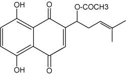
Acetyl shikonin
Because shikonin and its derivatives have biological activity against microorganisms, these compounds may play a role in plant defense in the rhizosphere. Several biotic factors (fungus, bacteria) increases the amount of total pigment, changes the ratios of derivatives produced, and initiates production of pigment de novo in epidermal cells (Brigham LA et al., Plant Physiol 1999, 119, 417). Shikonin derivatives function as both preformed and inducible microbial inhibitors regulated in a cell-specific manner to maximize the effect of highly toxic substances with minimal expense and harm to the plant.
Shikonin, extracted from the roots of another Boraginaceae, Lithospermum erythrorhizon (a Chinese herbal medicine with various antiviral and biological activities, including inhibition of human immunodeficiency virus type 1) has gained attention for its anticancer potential. Preclinical studies show that shikonin regulates multiple programmed cell death pathways, including apoptosis, necroptosis, ferroptosis, and pyroptosis. The molecular mechanisms and translational applications of that molecule have been reviewed (Lew CY et al., Nutrients 2025, 17(19), 3085).
Various analogues of shikonin have been isolated from a Boraginaceae, Alkanna cappadocica (Sevimli-Gur C et al., J Nat Prod 2010, 73, 860). They are all cytotoxic when tested on cancer cell lines.
More polar naphthoquinones have been shown to function in allelopathy as juglone or 5-hydroxy-1,4-naphthalenedione (Binder et al., Phytochemistry 1989, 28, 2799) and plant-insect interactions. It affects germination of plants less than it affects growth of the root and stem systems. Juglone is present in the leaves, roots, husks, fruit (the epicarp), and bark of plants of the Juglandaceae family (as walnut, Juglans regia). It is toxic to many plant species. It is sometimes used as an herbicide, a dye for cloth and inks, and a coloring agent for foods and cosmetics. Once exposed to air, the native and colorless hydroxyjuglone is degraded (oxydized) into juglone in changing its color from yellow to black.
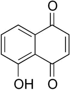 Juglone
Plumbagin is a yellow dye, named after the plant genus Plumbago, from which it was originally isolated. It is also commonly found in the carnivorous plant genera Drosera and Nepenthes. It is also a component of the black walnut drupe. It is regarded as a toxin which is genotoxic and mutagenic. The roots of Plumbago zeylanic have been used in Indian medicine for more than 2,500 years for treatments of various ailments. That compound has been shown to exert anticancer and antiproliferative activities in animal models and in cell culture (Aziz MA et al., Cancer Res 2008, 68, 9024).
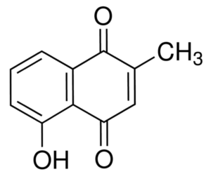 Plumbagin
Dehydro-alpha-lapachone is one of the numerous naphthoquinones isolated from several trees of the bignoniaceae family (Schmeda-Hirschmann et al., Z Naturforsch 2003, 58c, 495). The main source is the wood of Tabebuia heptaphylla, named “lapacho” in Paraguay. That wood is a traditional medicine used to treat wounds, inflammation and cancer (anti-vascularisation).
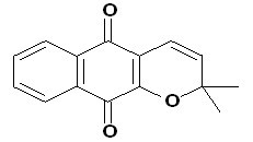
Dehydro-alpha-lapachone
Biological activities reported for lapachone comprises molluscicidal and trypanocidal effect. Lapachone can be considered a potential lead for the development of drugs to treat multidrug-resistant cell lines with lower expression of topoisomerase II (Krishnan P et al., Cancer Chemoth Pharm 2001, 47, 187). It was also shown to have antivascular activity in inhibiting vessel regeneration, interfering with vessel anastomosis (Schmeda-Hirschmann G et al., Z Naturforsch 2003, 58c, 495).
Seriniquinone is a bioactive natural product produced by the as-yet-undescribed marine bacterium of the rare marine bacterium of the genus Serinicoccus. That sulfur-containing compound has the unusual molecular formula C20H8O4S. It was discovered as a potential melanoma drug (Trzoss L et al., PNAS 2014, 111, 14687). Investigations revealed that seriniquinone has a remarkable in vitro activity against a diversity of cancer cell lines in the US National Cancer Institute 60-cell line screening. An overview of the discovery and development efforts to date are described in the Marine Drugs review (Hirata AS et al., Mar Drugs 2022, 20, 301).
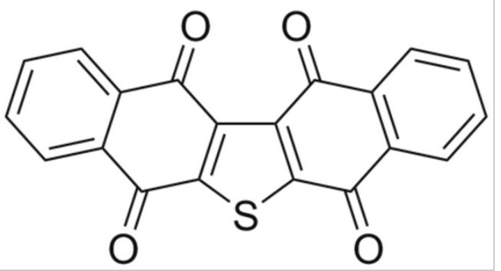 Seriniquinone
DISPERSIVE LIQUID-LIQUID MICROEXTRACTION
Lire la suiteDevenez membre et participez au développement de la Lipidomique au XXIème siècle.
S'inscrire