
QUANTIFICATION OF TRIACYLGLYCEROLS
BY HPLC
The quantitative determination of triacylglycerols in a sample can be made:
– after a previous separation by TLC
The spots are scraped and the lipids are eluted by washing 3 times the silica gel with 2 ml of dichloromethane or hexane/diethyl ether (1/1, v/v). After evaporation, dissolve the extract with a known volume of hexane/dichloromethane (9/1, v/v) before injection on the column.
– directly on the tissue extract
The tissue is preferably powdered in liquid nitrogen to prevent anay degradation. We extract by vortexing 10-15 min 500 mg of tissue powder (yet frozen) in a mixture of 3 ml ethanol, 2 ml water, 4 ml hexane and 4 ml diethyl ether.
After centrifugation, the upper phase is collected, the lower phase is washed with 8 ml hexane, the two upper phases are evaporated and the residue is dissolved in 2 ml dichloromethane. Dilute an aliquot with 10 volumes of hexane and inject 20-30 µl (about 5-10 ng of triacylglycerols) on the HPLC column.
Since the extract contains most often triacylglycerols but also cholesterol esters and cholesterol, we have devised a HPLC procedure using an isocratic condition to separate rapidly and if needed, to quantify these components.
HPLC separation
Apparatus
HPLC pump, injector
Evaporative Light Scattering detector (DDL 31, Eurosep)
Column: CN bonded phase, Merck (250 x 4.6 mm)
Reagents
Hexane, dichloromethane
Procedure
Prepare the mobile phase: mix 93 ml hexane with 7 ml dichloromethane.
The flow rate is 1 ml/min during 5 min to elute cholesterol esters and triacylglycerols, thereafter 3 ml/min during 10 min to elute cholesterol.
The evaporation temperature on the DDL is set at 50°C and the high voltage at 300V.
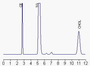
CE: cholesterol esters, TG: triacylglycerols, CHOL: cholesterol
The relationship between the amount of triacylglycerol injected and the peak area is not a straight line but can be linearized by logarithmic transformation of the triacylglycerol concentrations and the area units/1000 calculated by an electronic integrator. The calibration curve spreads over about two decades and the b value was 0.657 (r = 0.998). With a signal-to noise ratio of 3 , the detection limit of the detector is aboout 25 ng per injection. We use trilinoleine as standard.
A similar but simpler method has been described to determine the amount of triglycerides in diesel fuel using a silica column with an isocratic mobile phase (hexane and t-butyl ether) (Foglia TA et al., Chromatographia 2005, 62, 115). The method can also be sused for quantitating oils in petrodiesel.
An application note with details on the determination of triglycerides, diglycerides, and monoglycerides in biodiesel with a light scattering device (Corona Charged aerosol detector) may be found on the manufacturer web site.
DISPERSIVE LIQUID-LIQUID MICROEXTRACTION
Lire la suiteDevenez membre et participez au développement de la Lipidomique au XXIème siècle.
S'inscrire