DIACYLGLYCEROL
MOLECULAR SPECIES
After derivatization and purification by TLC, the fluorescent DAG are separated and quantified by HPLC on a C18 reversed-phase column (Lichrosphere 100RP-18, 5µ, Merck) at room temperature with acetonitrile/isopropanol (95/5, v/v) as mobile phase. The flow rate is 1 ml/min. The detection is made with a spectrofluorimeter (excitation: 230 nm, emission: 352 nm). We have used a thermostated column (13-15°C) to improve the separation of complex mixtures.
The mass proportion of each molecular species is calculated as the ratio of each peak area to the total surface of the detected peaks. With the use of an external standard, it becomes possible to determine the absolute mass of the each DAG injected. Sometimes, we used as an internal standard 14:1/14:1-DAG, this allows a direct correction for the recovery. The proposed method is very sensitive to picomole levels, the linearity of the response being verified from 15 pmol to 10 nmol of a single molecular species and the detection limit being at least 1 pmol per injection.
The identification of each peak is made by derivatization and HPLC analysis of commercial DAG standards, DAG mixtures formed by phospholipase C hydrolysis of known phospholipids and if necessary by collection of peaks and GLC analysis of the fatty acids in each fraction. The identification can also be made by developing a log RRT/carbon number plot as previously described (Nakagawa Y et al J Lipid Res 1983, 24, 1268).
|
|
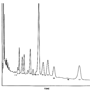
This chromatogram shows the separation of derivatized DAG from a rat brain phosphatidylethanolamine fraction. The main peaks correspond to: 4: 16:0/22:6, 5: 18:1/20:4 6: 16:0/20:4, 8: 18:0/22/6 11: 18:0/20:4, 1218:1/18:1 13: 16:0/18:1, 17: 18:0/18:1 It is assumed that the less saturated acyl group is at the 1-position. Thus 16:0/18:1 represents the 1-palmitoyl 2-oleoyl molecular species of a glycerophospholipid. |
|
|
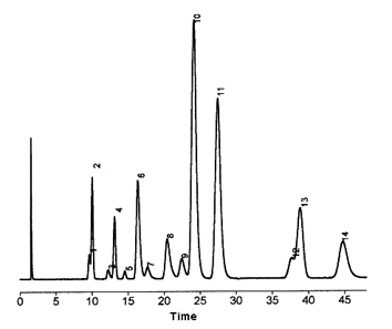
This chromatogram shows the separation of derivatized DAG obtained from rat brain phosphatidylcholine. The main peaks correspond to: 2: 16:0/22:6, 4: 16:0/20/4 8: 18:0/22:6, 10: 16:0/18:1 11: 16:0/16:0, 13: 18:0/18:1 14: 16:0/18:0 |
|
|
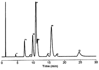
This chromatogram shows the separation of derivatized DAG molecular species obtained from a soja phosphatidylcholine fraction. The main peaks correspond to: 2: 18:2/18:3, 4: 18:2/18:2 5: 14:0/14:0 added as an internal standard 6: 16:0/18:3, 8: 16:0/18:2 10:18:0/18:2 |
DISPERSIVE LIQUID-LIQUID MICROEXTRACTION
Lire la suiteDevenez membre et participez au développement de la Lipidomique au XXIème siècle.
S'inscrire